45 compound microscope drawing with label
How to draw Microscope diagram for beginners - step by step How to draw Microscope diagram for beginners - step by step Perhaps Bidesh 52.4K subscribers Subscribe 5.3K 404K views 2 years ago Biology diagram Today I will show you " How to draw Microscope... Compound Microscope: Definition, Diagram, Parts, Uses, Working ... - BYJUS A compound microscope is defined as A microscope with a high resolution and uses two sets of lenses providing a 2-dimensional image of the sample. The term compound refers to the usage of more than one lens in the microscope. Also, the compound microscope is one of the types of optical microscopes.
1.5: Microscopy - Biology LibreTexts Labeled parts of a microscope. General Rules Always START and END with the low power lens when putting on OR taking away a slide. Never turn the nose piece by the objective lens. Do not get any portion of the microscope wet - especially the stage and objective lenses. Use only lens paper to clean microscope lenses. Cleaning the Microscope
Compound microscope drawing with label
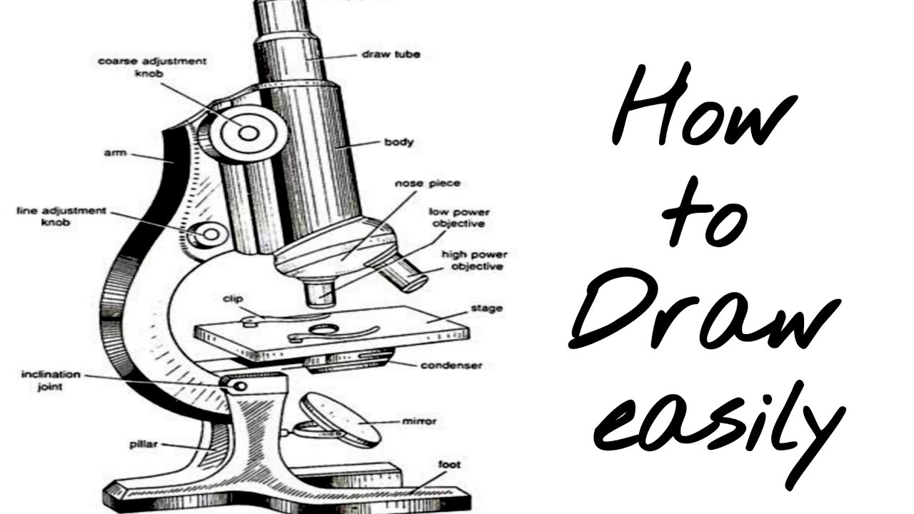
How to Draw a Microscope - Really Easy Drawing Tutorial Microscopes are used to investigate small objects that are too little to be seen with the naked eye. The type of microscope in our drawing guide is known as a compound microscope, an optical microscope, or a light microscope. It was first invented in the early 1600s. Parts of a microscope with functions and labeled diagram - Microbe Notes Parts of a microscope with functions and labeled diagram September 17, 2022 by Faith Mokobi Having been constructed in the 16th Century, Microscopes have revolutionalized science with their ability to magnify small objects such as microbial cells, producing images with definitive structures that are identifiable and characterizable. Exercise 1: Using a Compound Microscope | SpringerLink Procedure. 1. Retrieve the microscope from the cabinet by holding the arm and base of the microscope to provide support. 2. Take the microscope to your bench and plug it into the outlet on the side of the bench (some lab benches will have the plugs in the center of the bench). 3.
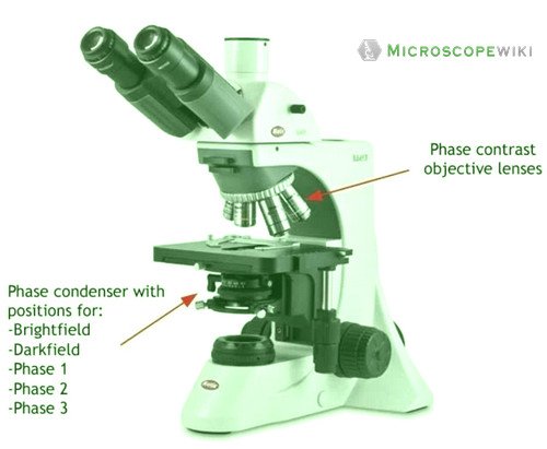
Compound microscope drawing with label. Labelled Diagram of Compound Microscope Labelled Diagram of Compound Microscope Article Shared by ADVERTISEMENTS: The below mentioned article provides a labelled diagram of compound microscope. Part # 1. The Stand: The stand is made up of a heavy foot which carries a curved inclinable limb or arm bearing the body tube. Microscope | Biology I Laboratory Manual - Lumen Learning Another microscope that you will use in lab is a stereoscopic or a dissecting microscope. This type of microscope uses visible light view thicker, larger specimens, such as an insect, in 3D. Since you are viewing larger samples, the magnification range of the dissecting microscope is lower than the compound light microscope. Compound Microscope: Parts of Compound Microscope - BYJUS The parts of the compound microscope can be categorized into: Mechanical parts; Optical parts (A) Mechanical Parts of a Compound Microscope. 1. Foot or base. It is a U-shaped structure and supports the entire weight of the compound microscope. 2. Pillar. It is a vertical projection. This stands by resting on the base and supports the stage. 3. Arm Compound Microscope Parts, Functions, and Labeled Diagram Compound Microscope Definitions for Labels Eyepiece (ocular lens) with or without Pointer: The part that is looked through at the top of the compound microscope. Eyepieces typically have a magnification between 5x & 30x. Monocular or Binocular Head: Structural support that holds & connects the eyepieces to the objective lenses.
Stock Images, Photos, Vectors, Video, and Music | Shutterstock Stock Images, Photos, Vectors, Video, and Music | Shutterstock Compound Microscope- Definition, Labeled Diagram, Principle, Parts, Uses Compound microscopes have a combination of lenses that enhances both magnifying powers as well as the resolving power. The specimen or object, to be examined is usually mounted on a transparent glass slide and positioned on the specimen stage between the condenser lens and objective lens. 3.8: Cell Structures and Organelles - Biology LibreTexts Using the compound microscope, look for small, red chromoplasts within the cells. Next, look for breaks in the regions between the cells. These are the plasmodesmata. Draw two adjacent pepper epidermal cells. Label the cell wall, middle lamella, plasmodesmata, and chromoplasts. You are encouraged to identify and label other cell components ... Compound Microscope Parts - Labeled Diagram and their Functions Labeled diagram of a compound microscope Major structural parts of a compound microscope There are three major structural parts of a compound microscope. The head includes the upper part of the microscope, which houses the most critical optical components, and the eyepiece tube of the microscope.
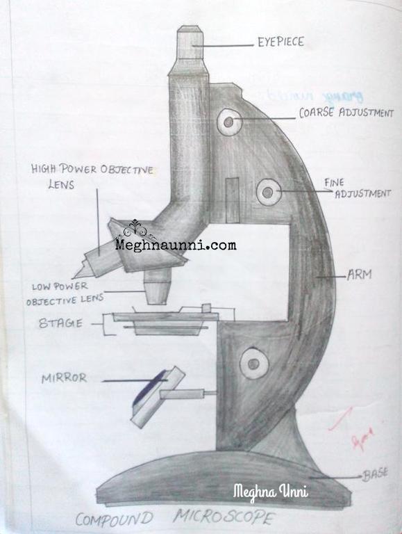
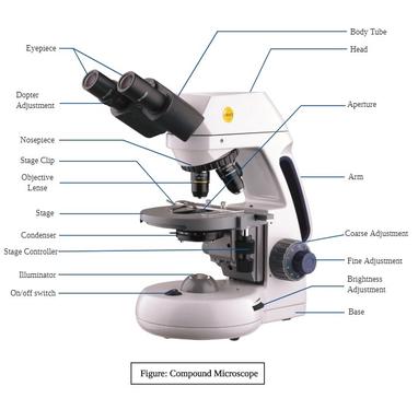
Microscope Parts and Functions Microscope Parts and Functions With Labeled Diagram and Functions How does a Compound Microscope Work? Before exploring microscope parts and functions, you should probably understand that the compound light microscope is more complicated than just a microscope with more than one lens. Compound Microscope - Diagram (Parts labelled), Principle and Uses Compound Microscope Parts (Labeled diagram) A compound microscope basically consists of optical and structural components. Within these two systems, there are multiple components within them and they are: Image : Labeled Diagram of compound microscope parts See: Labeled Diagram showing differences between compound and simple microscope parts Microscope Drawing - YouTube How to draw and label the Compound Microscope on Biology Note Book, Punjab Board Lahore by Naveed Akhtar Uppal(calligrapher & artist) Jhelum Label the microscope — Science Learning Hub In this interactive, you can label the different parts of a microscope. Use this with the Microscope parts activity to help students identify and label the main parts of a microscope and then describe their functions. Drag and drop the text labels onto the microscope diagram.
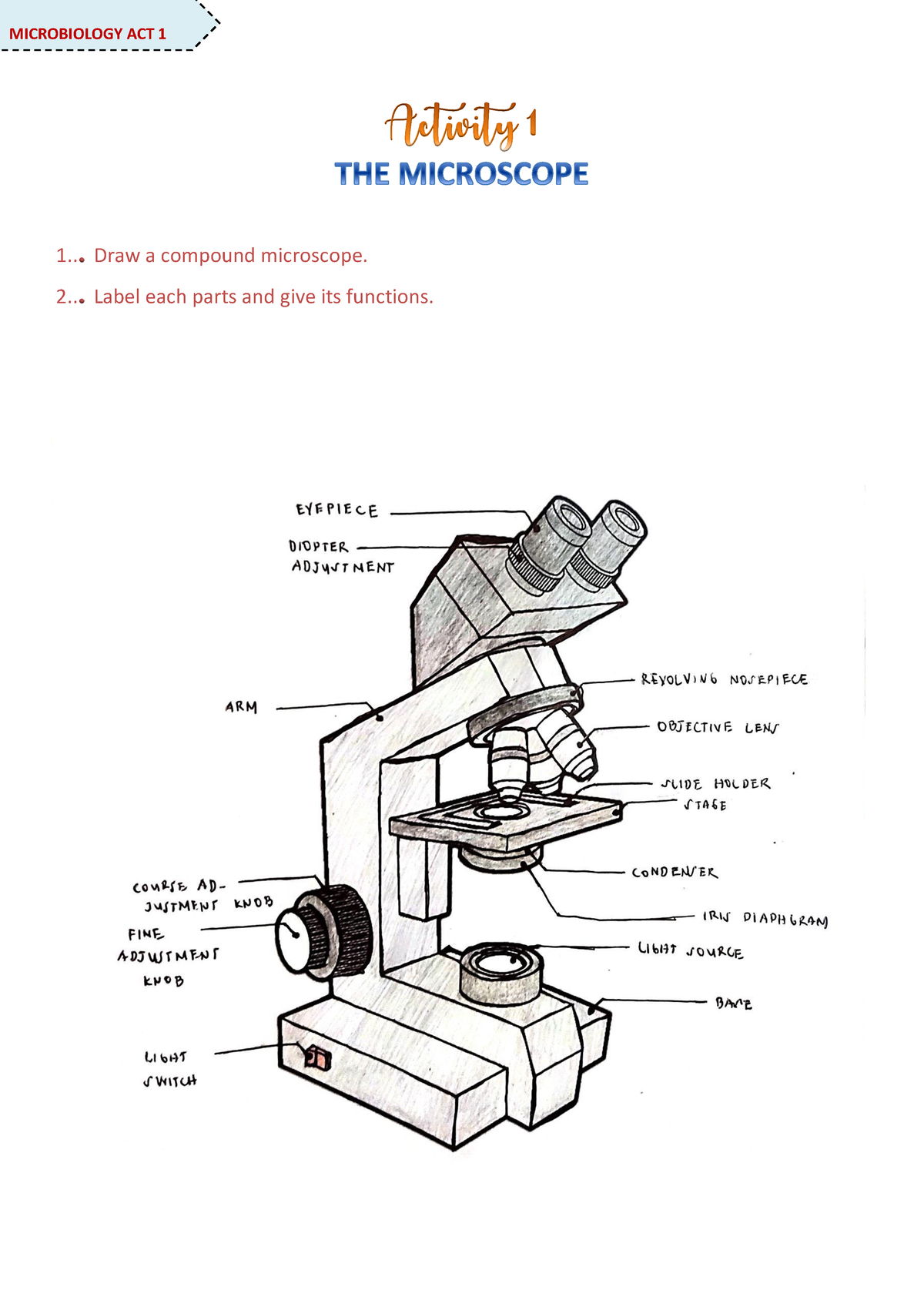
Microscope Activity - MICROBIOLOGY - 1... Draw a compound ...
How to draw compound of Microscope easily - step by step How to draw compound of Microscope easily - step by step Perhaps Bidesh 51.7K subscribers Subscribe 1.4M views 3 years ago Biology diagram I will show you " How to draw compound of microscope...
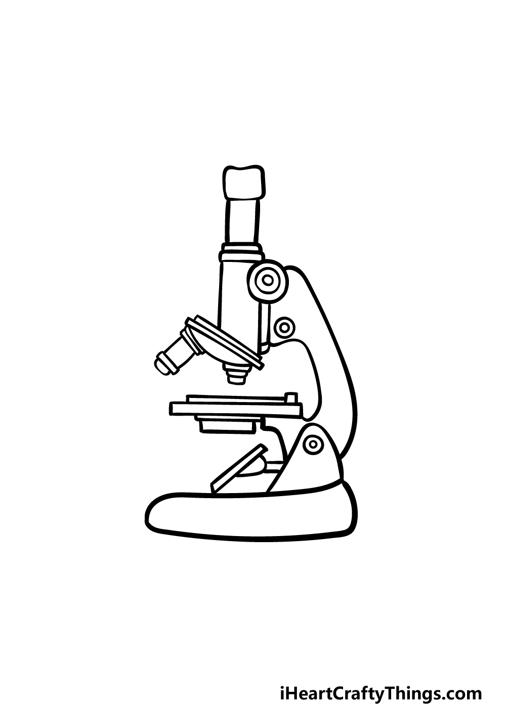
Microscope Drawing - How To Draw A Microscope Step By Step
Microscope Drawing Easy with Label - YouTube Microscope Drawing Easy with Label 898 views Apr 13, 2020 3 Dislike Share DrDiclonius 1.76K subscribers In this video I go over a microscope drawing that is easy with label. There is a...

How TO Draw microscope step by step easy/microscope drawing
Exercise 1: Using a Compound Microscope | SpringerLink Procedure. 1. Retrieve the microscope from the cabinet by holding the arm and base of the microscope to provide support. 2. Take the microscope to your bench and plug it into the outlet on the side of the bench (some lab benches will have the plugs in the center of the bench). 3.

Compound Microscope Parts, Function, & Diagram | What is a ...
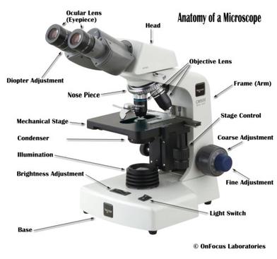
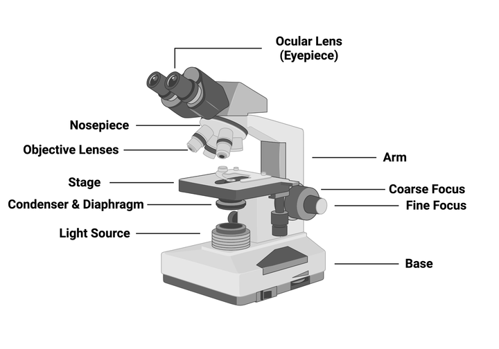
Parts of a microscope with functions and labeled diagram - Microbe Notes Parts of a microscope with functions and labeled diagram September 17, 2022 by Faith Mokobi Having been constructed in the 16th Century, Microscopes have revolutionalized science with their ability to magnify small objects such as microbial cells, producing images with definitive structures that are identifiable and characterizable.
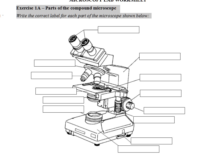
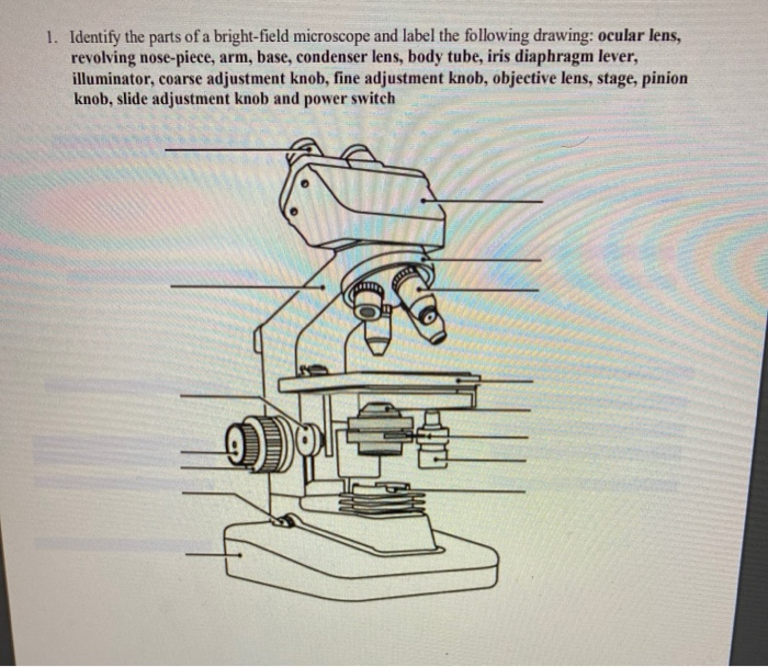
SOLVED: Exercise 1A Parts ofthe compound microscope Write the ...
How to Draw a Microscope - Really Easy Drawing Tutorial Microscopes are used to investigate small objects that are too little to be seen with the naked eye. The type of microscope in our drawing guide is known as a compound microscope, an optical microscope, or a light microscope. It was first invented in the early 1600s.
Biology : Compound Microscope Diagram for Class 8 ...

Parts of a microscope with functions and labeled diagram

HOw to draw light or compound microscope step by step / Microscope diagram
Solved 7. The Microscope 1. In a compound microscope: a. The ...

Draw a neat labelled diagram of a compound microscope. Derive ...
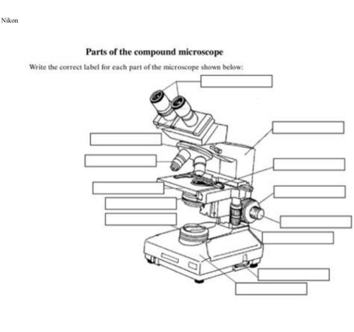
Solved Nikon Parts of the compound microscope Write the ...

Compound Microscope Parts, Functions, and Labeled Diagram ...

Collection Of Free Microscopes Drawing Label Clipart ...

I. Label the parts of a Compound Microscope. You may choose ...
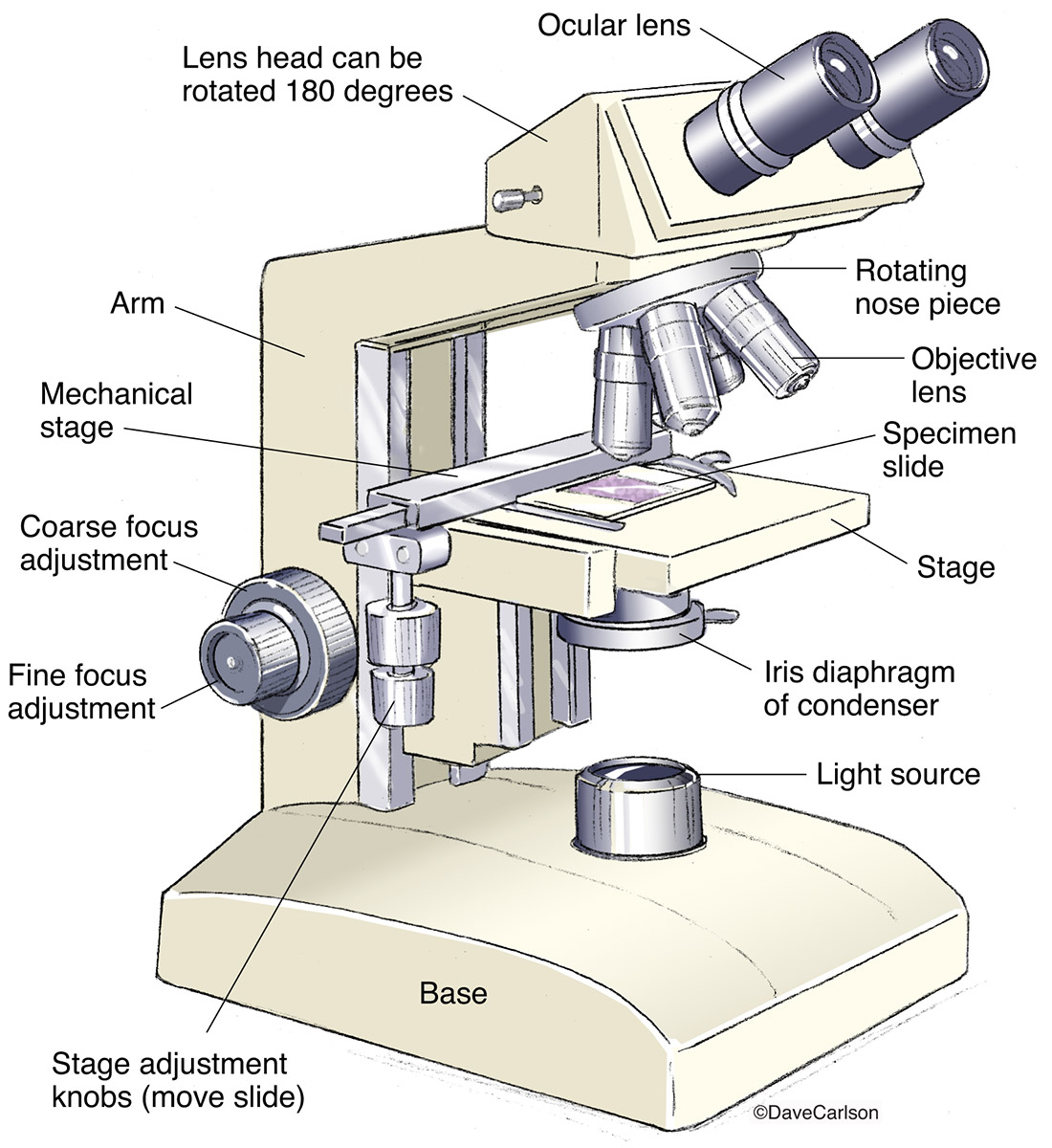
Compound Microscope | Image License | Carlson Stock Art
Microscope Parts and Functions

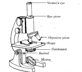
Simple Microscope - Parts, Functions, Diagram and Labelling ...
Microscope Types (with labeled diagrams) and Functions

Compound Microscope: Parts of Compound Microscope

Labelled Diagram of Compound Microscope | Figure Of Compound ...
How to draw compound microscope | science apparatus | compound microscope
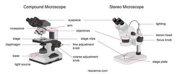
Parts of Stereo Microscope (Dissecting microscope) – labeled ...

Simple doodles, Microscopic images, Microscope parts
Compound Microscope Principle, Parts, Diagram Definition ...

Types, Parts and Functions of a Microscope
2.2 - Microscope Madness Workbook - kyoussef-mci

The compound light microscope
Microscope Clip Art at Clker.com - vector clip art online ...
Addgene: Using a Light Microscope Protocol

Can someone can send me diagram of this compound microscope ...
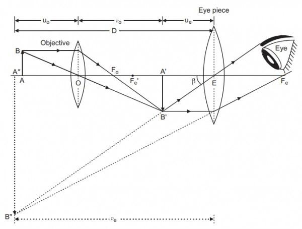
a) Draw a labeled ray diagram showing the formation of a ...
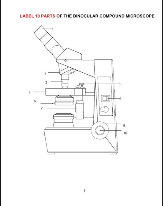
Solved LABEL 10 PARTS OF THE BINOCULAR COMPOUND MICROSCOPE ...

Simple Microscope - Diagram (Parts labelled), Principle ...

Compound Microscope Principle, Parts, Diagram Definition ...

The Microscope

label microscope diagram | Charts | Microscope, Anatomy bones ...

How to Draw a Microscope - Really Easy Drawing Tutorial

Microscope Diagram Labeled, Unlabeled and Blank | Parts of a ...

Microscope Parts, Types & Diagram | What is a Microscope ...

File:Labelledmicroscope.gif - Wikimedia Commons

Draw the structure of compound microscope - Brainly.in

Microscope label Diagram | Quizlet

in a long bond paper draw and label the parts of a compound ...
Draw a labelled diagram of a compound microscope.
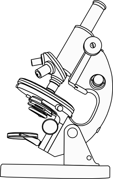
compound-microscope-unlabeled.jpg
Post a Comment for "45 compound microscope drawing with label"